Mohammad Badrul Anam
52nd annual meeting of the Japanese Society for Developmental Biologists (co-sponsored by Asia-Pacific Developmental Biology Network, held in Osaka International House, May 14th – 17th, 2019): An opportunity to share and exchange knowledge of advanced developmental biology studies
52nd annual meeting of the Japanese Society for Developmental Biologists (co-sponsored by Asia-Pacific Developmental Biology Network, held in Osaka International House, May 14th – 17th, 2019), was an amazing experience where scientists of corresponding fields from around the world presented their cutting edge research findings. It was a great opportunity to share my ongoing research findings as well as exchange knowledge of advanced techniques to hone our research skills.
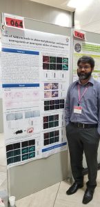
I participated last year in this conference, but this time it was fascinating for me as I have given an oral scientific research presentation along with poster presentation for the first time in an international level. There I presented my research work titled “Loss of Akhirin leads to abnormal phenotype and impaired neurogenesis at neurogenic niches of mouse brain”. Akhirin (AKH) is a secreted protein that was first identified in embryonic chicken eye (Ahsan et al, 2004) development. Later its involvement was reported on the development of mouse spinal cord and its maintenance activity following an induced spinal cord injury (Athary et al, 2014). AKH is a secreted protein comprised of 748 amino acid including one LCCL domain at its N-terminal and two von Willebrand factor A (VWA) at its C-terminal. LCCL implies the Limulus factor C (a serine protease), Coch 5b2 (found in inner ear and mutation in DNA9 causes deafness) and late gestational lung protein 1 or Lgl1 is involved in fetus lung development and associated with Limulus factor C and Coch 5b2. VWA interacts with platelets and collagen in blood clotting event. Now our lab is interested to explore its function in two zones of mouse brain that harbors stem cells i.e. lateral ventricle (LV) lining the subventricular zone (SVZ) and dentate gyrus (DG) of hippocampus. In my presentations (both oral and poster) I have shown some of the primary experimental data observed on AKH. Already By in situ hybridization assay, I have found a robust expression pattern of AKH at E17 (Embryonic day-17)and P0 (Post-natal day-0) SVZ and DG regions which are subsequently downregulated as development proceeds. At P20 the signal of AKH is completely faded away in SVZ and DG but remains restricted at CA2 regions of hippocampus. CA2 region is thought to be involved in social-cognitive learning process. This result indicate AKH might have role localizing in the neurogenic niches of mouse brain. By nissl staining we have observed that LV area is significantly increased in AKH knockout mice (AKH-/-) but DG area is decreased than its wild type (WT) counterpart. And a few population (3%) has developed hydrocephalus type phenotype. These results indicate loss of AKH induces defective developmental phenotype at neurogenic niches. Next we performed BrdU staining that showed reduced BrdU+ cells in both AKH-/- SVZ and DG areas. This indicates AKH is involved in cell proliferation at neurogenic niches. Also we performed triple staining by GFAP (neural stem cell marker), SOX2 (Neural stem cell/progenitors marker) and Ki67 (Cell proliferation marker) and found that Ki67+ cells, GFAP+/SOX2+/Ki67+ cells reduced significantly in AKH-/- DG. That indicates cell proliferation and proliferating neural stem cell (NSC) have been reduced in the DG. Also quiescent neural stem population increased slightly by observing GFAP+/SOX2+ cells. However, we have not performed this triple staining in SVZ so we cannot conclude whether it has similar effects in SVZ or not. Neither we can conclude the increased population quiescent NSCs. We have also observed smaller sized neurospheres from SVZ and hippocampal regions of AKH-/- and that indicates lack of AKH may hamper the regulation of NSCs. We performed immunohistochemical (IHC) assay on SVZ-neuropheres by Nestin (neural stem cell/progenitor cell maker) and Ki67 and found that both marker’s signal has been reduced. However we have not performed this investigation on DG-neurospheres but we are expecting same result from it. This indicates due to reduction of cell proliferation the size of the neurospheres has become reduced. We also performed In vitro differentiation experiment from these neurospheres and labeled the differentiated cells by Tuj1 (immature neuronal marker) though IHC. We found that Tuj1+ cells and length of neurites have been reduced significantly from both AKH-/- SVZ and DG neuropheres differentiated cells. This indicates loss of AKH impairs the NSCs differentiation. All together my experimental data has suggested that AKH has a pivotal role localizing at neurogenic niches and regulates the NSCs proliferation and differentiation in mouse brain.
During and after the presentations I have got numbers of appreciations and many of the appreciators were expert on this filed and gave me some valuable suggestion and I discussed with them to improve my experimental directions. For example one of the audiences indicates that it shows some heterophilic cell-adhesion property, so how its mechanism of action can be addressed in correlation with that property while interacting with other signaling molecules. Another audience comments on investigating the specific markers on hydrocephalic brain. Also I have got suggestions about investigating behavioral anomalies of AKH-/- mice as because we have observed loss of AKH showed reduced differentiation potentiality than WT. This investigation is already in cue to perform soon. Some experts also suggested me to investigate more in SVZ NSCs regulation. Another audience also suggested about role of AKH on traumatic brain injury model as in previous report AKH showed some functions on mouse spinal cord injury model. This can be investigated as a plan for future project of AKH. So overall the appreciations and the suggestions of the audiences will encourage us to perform more extensive research on AKH in future.
Besides my presentation session I was fascinated by the works of Ms. Munkhsoyol Erkhembaatar on “The role of the Strawberry Notch Homolog 1 in the neurite growth of the cortical neurons”. There she investigated the loss of function of SBNO1, a mammalian homologue of Drosophila gene Strawberry Notch. The conditional knockout (KO) SBNO1 mice showed drastically reduced brain morphogenesis that the WT type mice. The cerebral cortex was abnormally thinner and SBNO KO mice were severely paralyzed. In the brain stem the pyramidal tracts of neurons were consistently absent. This is an interesting study and I discussed with her if she had performed In vitro differentiation experiment as because I guessed SBNO1 is affecting neuronal regulation as like in my study with AKH. This experiment can possibly give a strong supportive data of her in vivo results. By this discussion she appreciated the concept she has got from AKH’s in vitro experimental result.
Later another presentation attracted me which was presented by Ms. Mai Ahmed from Okinawa Institute of Science and Technology (“Mutation in strip1 gene leads to impaired retinal neural
circuit formation in zebrafish.”) where she showed rw147 mutant zebra fish showed lamina developmental defects where missense mutation was occurred in striatin-interacting protein 1 (strip1). She also suggested an impaired localization of retinal cells type was occurred due to the mutation where inner nuclear layer cells (INL) was wrongly positioned in ganglion cell layer (GCL). Bipolar cells of mutant zebra fish showed disturbed neurite extension patterns and defects in branching through mosaic labeling. In this I also found interest and discuss with her about AKH’s role in chicken lens and retina development. We discussed as strip1 is enriched in plasma membrane and AKH also has heterophilic cell adhesion property so there can be an interaction between these two molecules in cell-cell communication. As because currently she is working with zebra fish, Therefore investigation of interaction between AKH and strip1 can be a future research project on mammals.
Besides my field of interest another presentation in the conference has caught my attention. The presentation titled “A signal mediated by retinoic acid functions as a novel regulative step for allowing zebrafish fin regeneration” was presented by Dr. Atsushi Kawakami. There he showed that retinoic acid (RA) pathway is critical in the regulation of regeneration process on zebra fish fin. He did not mentioned the name but used a retinoic acid receptor agonist that impaired the regeneration process and induced cyp26a (the enzyme for retinoic acid degradation) expression strongly. There he concluded balance between RA and cyp26a is critical in regeneration process and this event can be a regulatory factor for regenerative capacity. In our discussion I have asked him whether he has investigated in other species (specially in mammals). But his works are currently based on zebra fish but he is interested to observe similar effects in other species. In my assumption if his study could make a breakthrough in this field, it will certainly make a great contribution in regenerative medicine.
Finally, I would like to thank my lab Department of Developmental Neurobiology where everybody helped me to get experienced in an international scientific environment like JSDB.
