


Yohei Sasagawa, Kunitoshi Yamanaka, Shingo Nishikori, and Teru Ogura (2007) Caenorhabditis elegans p97/CDC-48 is crucial for progression of meiosis I. Biochem. Biophys. Res. Commun. 358, 920-924.
p97/VCP/Cdc48p belongs to the AAA (ATPases associated with diverse cellular activities) family and has been indicated to be required for mitotic M-phase. We previously reported that simultaneous depletion of two p97 homologues, CDC-48.1 and CDC-48.2, in Caenorhabditis elegans caused the complete embryonic lethality, and that a large number of vacuole-like structures were observed in the dead embryos. However, cellular functions of p97 in embryogenesis have not been revealed. In this study, we analyzed effects of p97 depletion on meiotic progression. Simultaneous depletion of both p97 resulted in the formation of aberrant multinucleate cells and sometimes ectopic furrows in embryos. Importantly, meiotic chromosomes were not divided at meiotic metaphase I in p97-depleted embryos, although spindle formation and disassembly occurred. Furthermore, we found that chromosome condensation was significantly reduced in p97-depleted oocytes. Taken these results altogether, we propose that C. elegans p97 plays an important role in the progression of meiosis.
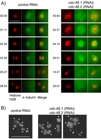
A Requirement of p97 for progression of meiotic metaphase I. CDC-48.1 and CDC-48.2 were simultaneously depleted on OD57 for 24 hours at 20°C. Subsequently plates were incubated for additional 12 hours at 25°C to express GFP-fused α-tubulin, because the expression of the fusion protein is suppressed at 20°C. mCherry::Histone H2B (left), GFP::α-tubulin (middle) and merged (right) images were presented at each time point indicating minutes:seconds to the left). Scale bar: 10 μm in all images.
B Effect of p97 depletion on chromosome condensation in meiotic prophase I. Both p97 were depleted on wild-type N2 strain for 60 hours from L4 stage. Gonads were dissected, fixed and stained with DAPI. Asterisks show bivalents in maturated oocytes. White arrowhead represents an abnormal chromosome bridge. Bar represents 10 μm.