Soichiro Ogaki, Nobuaki Shiraki, Kazuhiko Kume, and Shoen Kume*,
* Corresponding author.
Wnt and Notch signals guide embryonic stem cell differentiation into the intestinal lineages, Stem Cells , 2013
The studies of differentiation of mouse or human embryonic stem (ES) cells into specific cell types of the intestinal cells would provide insights to the understanding of intestinal development and ultimately yield cells for the use in future regenerative medicine. Here, using an in vitro differentiation procedure of pluripotent stem cells into definitive endoderm, inductive signals pathways guiding differentiation into intestinal cells was investigated. We found that activation of Wnt/ b -catenin and inhibition of Notch signaling pathways, by simultaneous application of 6-bromoindirubin-3′-oxime (BIO), a glycogen synthase kinase ( GSK)-3 b inhibitor , and DAPT, a known γ-secretase inhibitor, efficiently induced intestinal differentiation of ES cells cultured on feeder cell. BIO and DAPT patterned the definitive endoderm (DE) at graded concentrations. Upon prolonged culture on feeder cells, all four intestinal differentiated cell types, the absorptive enterocytes and three types of secretory cells (goblet cells, enteroendocrine cells, Paneth cells), were efficiently differentiated from mouse and human ES cell-derived intestinal epithelium cells. Further investigation revealed that in the mouse ES cells, fibroblast growth factor (FGF) and bone morphogenetic protein (BMP) signaling act synergistically with BIO and DAPT to potentiate differentiation into the intestinal epithelium. However, in human ES cells, FGF signaling inhibited, and BMP signaling did not affect differentiation into the intestinal epithelium. We concluded that Wnt and Notch signaling function to pattern the anterior-posterior axis of the DE and control intestinal differentiation.

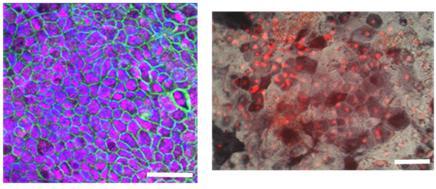
Fig. (Left) Mouse intestinal epithelial (IE) cells expressing an IE marker, Cdx2 (red), and an epithelial marker, E-cadherin (green) are shown. (Right) CDX2-positive human IE cells (red) exhibiting ALP activities (black) (ALP+/ CDX2+ = 79%) are shown.