


Mariam C. Recuenco*, Tomoko Ohmori*, Shunsuke Tanigawa, Atsuhiro Taguchi, Sayoko Fujimura, Mary Anne Conti, Qize Wei, Hiroshi Kiyonari, Takaya Abe, Robert S. Adelstein and Ryuichi Nishinakamura (2014) Non-muscle myosin II regulates the morphogenesis of metanephric mesenchyme-derived immature nephrons. J. Am. Soc. Nephrol. Epub ahead of print
* These authors contributed equally to this work.
The kidney develops from reciprocal interactions between the metanephric mesenchyme and ureteric bud. The mesenchyme transforms into epithelia and forms complicated nephron structures, whereas the ureteric bud extends its pre-existing epithelial ducts. Although the roles are well established for extracellular stimuli, such as Wnt and Notch, it is unclear how the intracellular cytoskeleton regulates these morphogenetic processes. Myh9 and Myh10 encode nonmuscle myosin II heavy chains, and Myh9 mutations in humans are implicated in congenital kidney diseases and focal segmental glomerulosclerosis in adults. Here, we analyzed the roles of Myh9 and Myh10 in the developing kidney. Ureteric bud-specific depletion of Myh9 resulted in no apparent phenotypes, whereas mesenchyme-specific Myh9 deletion caused proximal tubule dilations and renal failure (Figure: upper panels). Mesenchyme-specific Myh9/Myh10 mutant mice died shortly after birth and showed a severe defect in nephron formation. The nascent mutant nephrons failed to form a continuous lumen (Figure: lower panels), which likely resulted from impaired apical constriction of the elongating tubules. In addition, nephron progenitors lacking Myh9/Myh10 or the possible interactor Kif26b were less condensed at midgestation and reduced at birth. Taken together, nonmuscle myosin II regulates the morphogenesis of immature nephrons derived from the metanephric mesenchyme and the maintenance of nephron progenitors. Our data also suggest that Myh9 deletion in mice results in failure to maintain renal tubules but not in glomerulosclerosis.
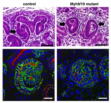
Figure
Morphological defects of mouse nascent nephrons in the absence of non-muscle myosin II
(Upper panels) While S-shaped immature nephrons (arrows) are observed in the control, Myh9/10 mutant nephrons are irregularly shaped.
(Lower panels) The aPKC-positive domain (red) delineates a single continuous lumen in the control, whereas the mutant lumina are discontinuous and branch irregularly. NCAM is stained green to visualize nascent nephrons.