


Shinya Sugimoto, Ken-ichi Okuda, Reina Miyakawa, Mari Sato, Ken-ichi Arita-Morioka, Akio Chiba, Kunitoshi Yamanaka, Teru Ogura, Yoshimitsu Mizunoe, Chikara Sato.
Imaging of bacterial multicellular behaviour in biofilms in liquid by atmospheric scanning electron microscopy.
Scientific Reports 6:25889 (2016).
doi:10.1038/srep25889
Biofilms are complex communities of microbes that attach to biotic or abiotic surfaces causing chronic infectious diseases. Within a biofilm, microbes are embedded in a self-produced soft extracellular matrix (ECM), which protects them from the host immune system and antibiotics. The nanoscale visualization of delicate biofilms in liquid is challenging. Here, we develop atmospheric scanning electron microscopy (ASEM) to visualise Gram-positive and -negative bacterial biofilms immersed in aqueous solution. Biofilms cultured on electron-transparent film were directly imaged from below using the inverted SEM, allowing the formation of the region near the substrate to be studied at high resolution (Figure 1). We visualized intercellular nanostructures and the exocytosis of membrane vesicles (Figure 2A, B), and linked the latter to the trafficking of cargos, including cytoplasmic proteins and the toxins hemolysin and coagulase. A thick dendritic nanotube network was observed between microbes, suggesting multicellular communication in biofilms. A universal immuno-labelling system was developed for biofilms and tested on various examples, including S. aureus biofilms. In the ECM, fine DNA and protein networks were visualized and the precise distribution of protein complexes was determined (e.g., straight curli, flagella, and excreted cytoplasmic molecular chaperones) (Figure 2C, D). Our observations provide structural insights into bacteria-substratum interactions, biofilm development and the internal microbe community.
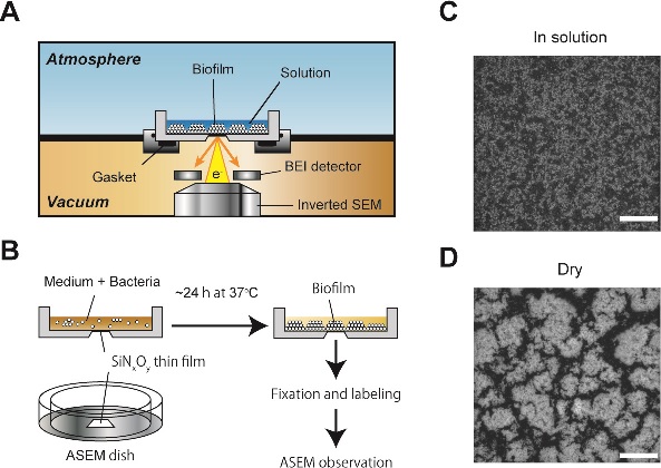
Figure 1. Outline of ASEM observation and visualization of soft biofilms. (A) Diagram of the ASEM showing the inverted SEM, the detector and the specimen dish, which separates the atmosphere (above) and the column vacuum (below). The electron beam passes through the window and is projected up onto the biofilms immersed in solution, penetrating them to a depth of 2-3 μm. Backscattered electrons (BSE) are captured by a BSE imaging (BEI) detector. (B) Bacteria were grown in an appropriate medium in the removable, 35-mm ASEM dish with SiNxOy film window at the center of its base. Various procedures labeling particular molecules are applicable. (C) Biofilms in solution. (D) Dried biofilms. Scale bars, 50 μm.
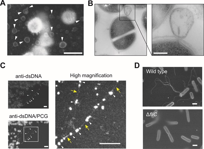
Figure 2. ASEM images of biofilms. (A) Heavy metal staining of S. aureus biofilms. Arrowheads indicate spherical structures. (B) Production of membrane vesicles (MVs) within biofilms of S. aureus observed by thin-sectioning TEM. (C) Biofilms of S. aureus were labeled with anti-dsDNA mouse IgG primary antibody and colloidal gold-conjugated anti-mouse IgG secondary antibody (anti-dsDNA). The biofilms were subsequently counter-stained with positively charged Nanogold (anti-dsDNA/PCG). A higher magnification image of the white rectangle is shown on the right. Arrowheads and arrows mark linearly aligned colloidal gold particles and PCG-positive fibrillary structures, respectively. (D) Visulalisation of E. coli flagella by PCG-labeling ASEM. E. coli wild-type and ΔfliC mutant, which lacks a gene involved in the biosynthesis of flagella, were labeled with PCG and observed in liquid by ASEM. Spiral flagella were prominent in the wild type. Scale bars, 1 μm (A, C, D) and 100 nm (B).