


Yoshikazu Hirate, Shino Hirahara, Ken-ichi Inoue, Atsushi Suzuki, Vernadeth B. Alarcon, Kazunori Akimoto, Takaaki Hirai, Takeshi Hara, Makoto Adachi, Kazuhiro Chida, Shigeo Ohno, Yusuke Marikawa, Kazuki Nakao, Akihiko Shimono, Hiroshi Sasaki
“Polarity-Dependent Distribution of Angiomotin Localizes Hippo Signaling in Preimplantation Embryos”
Current Biology (2013), http://dx.doi.org/10.1016/j.cub.2013.05.014
Background: In preimplantation mouse embryos, the first cell fate specification to the trophectoderm or inner cell mass occurs by the early blastocyst stage. The cell fate is controlled by cell position-dependent Hippo signaling, although the mechanisms underlying position-dependent Hippo signaling are unknown.
Results: We showed that a combination of cell polarity and cell–cell adhesion establishes position-dependent Hippo signaling, where the outer and inner cells are polar and nonpolar, respectively. The junction-associated proteins angiomotin (Amot) and Amotl2 are essential for Hippo pathway activation and appropriate cell fate specification. In the nonpolar inner cells, Amot localizes to adherens junctions (AJs) and cell–cell adhesion activates the Hippo pathway. In the outer cells, the cell polarity sequesters Amot from basolateral AJs to apical domains, thereby suppressing Hippo signaling. The N-terminal domain of Amot is required for actin binding, Nf2/Merlin-mediated association with the E-cadherin complex, and interaction with Lats protein kinase. In AJs, Ser176 in the N-terminal domain of Amot is phosphorylated by Lats, which inhibits the actin-binding activity, thereby stabilizing the Amot–Lats interaction to activate the Hippo pathway.
Conclusion: We propose that the phosphorylation of S176 in Amot is a critical step for activation of the Hippo pathway in AJs and that cell polarity disconnects the Hippo pathway from cell–cell adhesion by sequestering Amot from AJs. This mechanism converts positional information into differential Hippo signaling, thereby leading to differential cell fates.
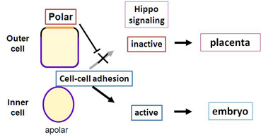
Figure legend
Combination of cell polarity and cell-cell adhesion controls Hippo signal
In preimplantation embryos, cell-cell adhesion activates the Hippo signal in the inner cells. In the outer cells, presence of cell polarity suppresses activation of the Hippo signal by cell-cell adhesion.