


Maki Takagishi, Binta Maria Aleogho, Masako Okumura, Kaori Ushida, Yuichiro Yamada, Yusuke Seino, Sayoko Fujimura, Kaoru Nakashima, and Asako Shindo*
Nutritional control of thyroid morphogenesis through gastrointestinal hormones.
Current Biology
DOI: 10.1016/j.cub.2022.01.075.
The thyroid gland secretes hormones that regulate metabolism and growth, and the endocrine function is based on a spherical tissue called the follicle. Some thyroid diseases show abnormalities in the morphology and size of the follicles. The molecular mechanism of controlling thyroid follicles morphology has been one of the most important medical and biological issues.
Using an amphibian model animal, African clawed frog (Xenopus laevis), the Laboratory of Morphogenesis (Associate professor Asako Shindo) has discovered that thyroid morphogenesis is regulated by external nutrients. Unlike mammals, which receive nutrients from the mother via the placenta, the Xenopus tadpoles eat during organogenesis to continue development. Three-dimensional images of Xenopus thyroid tissue at a cellular resolution revealed that nutrients from the food were necessary to initiate the formation of thyroid follicles. The unfed tadpoles did not start thyroid follicle formation, although the tadpoles exhibited normal swimming behavior and survived for a certain period of time (Figure, top).
Furthermore, the authors found that the gastrointestinal hormone, incretin, is necessary for the formation of the nutrients-dependent thyroid follicles formation. Analysis of the positional relationship between thyroid cells and the follicular lumina showed that incretin promotes connecting the multiple small follicles (schematic diagram, bottom). In addition, knockout mice of the receptor for GIP (Glucose-dependent insulinotropic polypeptide), one of the incretins, showed smaller thyroid follicles in neonates, indicating that the function of incretin in thyroid morphogenesis may be conserved across species.
This study indicates that developing animals have a system to changes organ morphology flexibly in response to nutritional status. Further clarification of the roles of nutrition and hormones during organogenesis may contribute to the elucidation of congenital thyroid hypoplasia and the mechanisms that maintain homeostasis of thyroid follicle morphology.
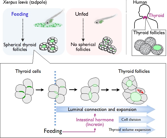
Schematic of the findings: Feeding initiates the thyroid follicle formation via gastrointestinal hormone.
Xenopus laevis tadpole was used as a main model. Feeding stimulates incretin secretion from the gastrointestinal tract, which in turn initiates and promotes thyroid follicle formation.