


Fahim Haque, Yusuke Kaku, Sayoko Fujimura, Tomoko Ohmori, Robert S. Adelstein, and Ryuichi Nishinakamura. Non-muscle Myosin II Deletion in the Developing Kidney Causes Ureter-bladder Misconnection and Apical Extrusion of the Nephric Duct Lineage Epithelia. Developmental Biology 427: 121-130, 2017.
In kidney development, connection of the nephric duct (ND) to the cloaca and subsequent sprouting of the ureteric bud (UB) from the ND are important for urinary exit tract formation. Although the roles of Ret signaling are well established, it remains unclear how intracellular cytoskeletal proteins regulate these morphogenetic processes. Myh9 and Myh10 encode two different non-muscle myosin II heavy chains, and Myh9 mutations in humans are implicated in congenital kidney diseases. Here Fahim Haque (graduate student of the HIGO program) and the members of Department of Kidney Development (Prof. Ryuichi Nishinakamura) report that ND/UB lineage-specific deletion of Myh9/Myh10 in mice caused severe hydroureter/hydronephrosis at birth (Figure A). At mid-gestation, the mutant ND/UB epithelia exhibited aberrant basal protrusion and ectopic UB formation (Figure B), which likely led to misconnection of the ureter to the bladder. In addition, the mutant epithelia exhibited apical extrusion followed by massive apoptosis in the lumen (Figure C), which could be explained by reduced apical constriction and intercellular adhesion mediated by E-cadherin. These phenotypes were not ameliorated by genetic reduction of the tyrosine kinase receptor Ret. In contrast, ERK was activated in the mutant cells and its chemical inhibition partially ameliorated the phenotypes. Thus, myosin II is essential for maintaining the apicobasal integrity of the developing kidney epithelia independently of Ret signaling.
Figure
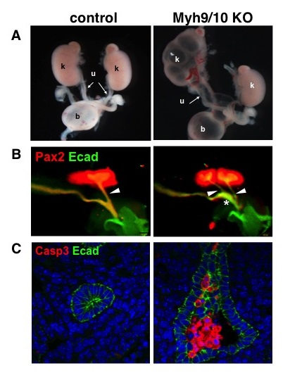
Abnormalities of the mouse kidneys in the absence of non-muscle myosin II
A.Hydronephrosis (k) and hydroureter (u) in the mutant mice at birth. Left panel: control mouse. b: urinary bladder.
B.The nephric duct is dilated (*) and multiple ureteric buds (arrowheads) are formed in mutant kidney (embryonic day 11.5).
C.The nephric duct cells are apically extruded from the epithelial layer (green: E-cadherin) and undergo apoptosis (red: cleaved caspase-3) in the mutant kidney (embryonic day 11.5).