


Our major research areas are muscle stem cell biology, muscle plasticity, muscle metabolism, and muscle diseases including muscular dystrophies and age-related sarcopenia. Our research projects are supported by the Program for Technological Innovation of Regenerative Medicine (AMED, Japan Agency for Medical Research and Development), the Practical Research Project for Rare/Intractable Diseases (AMED), Grants-in-Aid for Scientific Research (JSPS, Japan Society for the Promotion of Science), Center for Metabolic Regulation of Healthy Aging (Kumamoto University Faculty of Life Sciences), and International Research Core for Stem Cell-based Developmental Medicine (Kumamoto University Research Project).
Molecular regulation of muscle stem cell fate decisions
Satellite cells are the resident muscle stem cells that are located between the basal lamina and the plasmalemma of myofibers, providing new myonuclei for postnatal muscle growth and hypertrophy, and regeneration in adult muscle (Figure 1). Satellite cells have a potent regenerative ability and thus they hold promise for treating muscle weakness such as muscular dystrophy and sarcopenia.
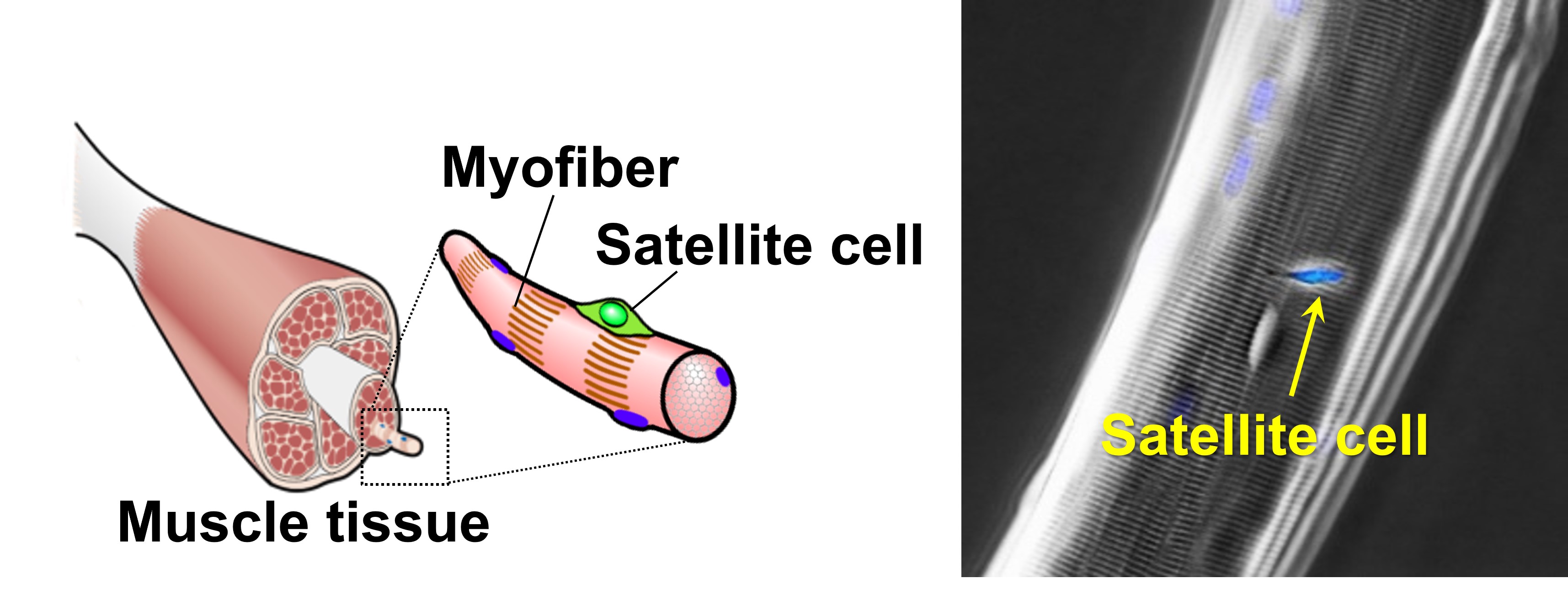
Figure 1. Muscle stem cell (Satellite cell)
In healthy adult muscle, satellite cells are mitotically quiescent but they are activated in response to stimulation such as muscle injury. Activated satellite cells proliferate extensively and the majority of proliferated cells then undergo myogenic differentiation to form myofibers, while the others return to a quiescent state to self-renew and maintain the stem cell pool. Our laboratory works to understand molecular mechanisms that control the satellite cell fates of proliferation, differentiation or self-renewal (Ono et al., J Cell Sci 2009; Ono et al., Cell Death Differ 2011; Ono et al., Cell Rep 2015; Fujimaki et al., Stem Cells 2018)(Figure 2).
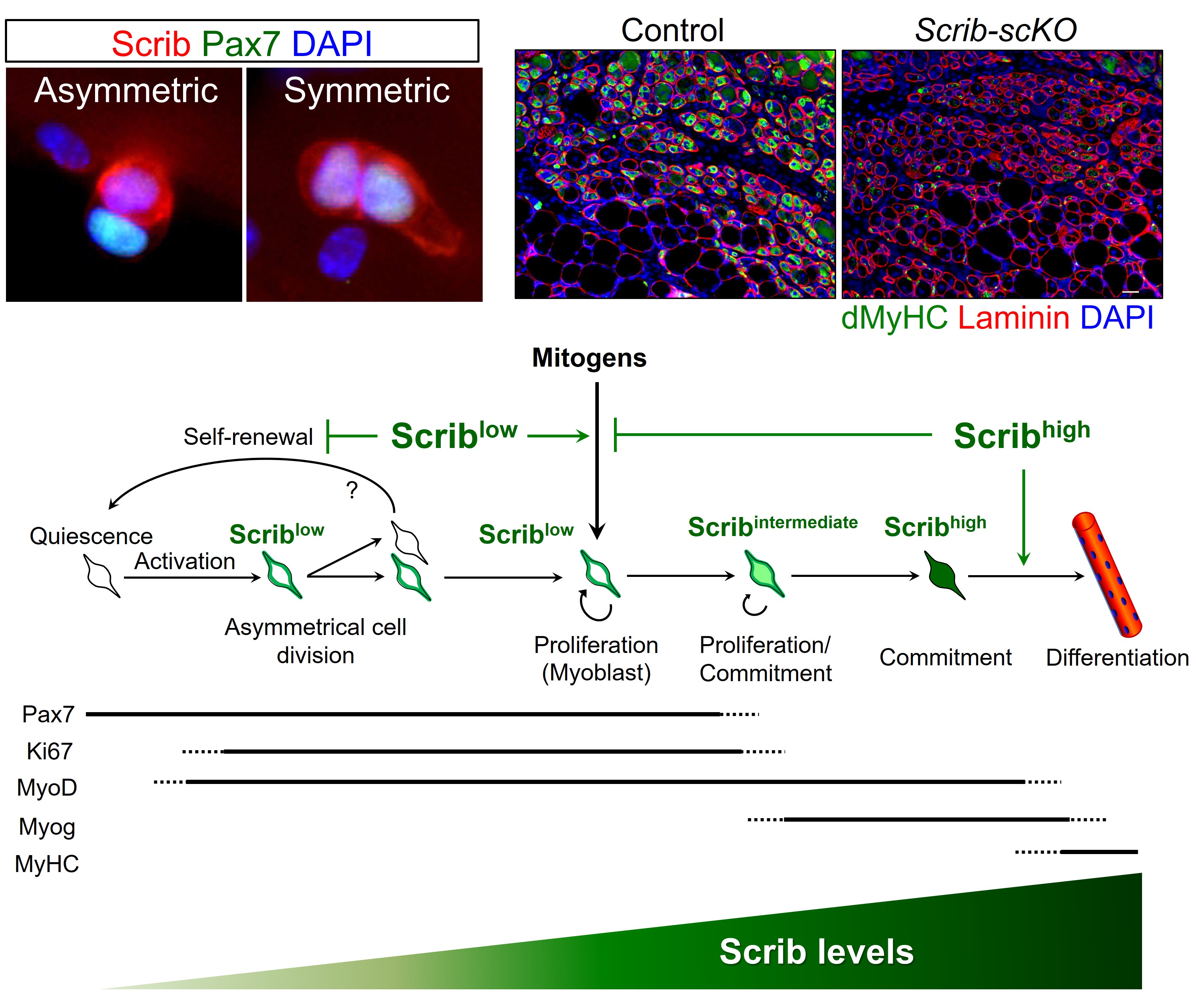
Figure 2. Scrib dictates satellite cell fates
We have also generated a knock-in mouse line expressing a Pax7-enhanced yellow fluorescent protein (YFP) fusion protein that allows us to visualize living satellite cells through Pax7-YFP fluorescence (Kitajima and Ono, Skelet Muscle 2019)(Figure 3). Using this newly established mouse line, we are undertaking to develop an in-vitro tissue-engineered muscle regeneration model.
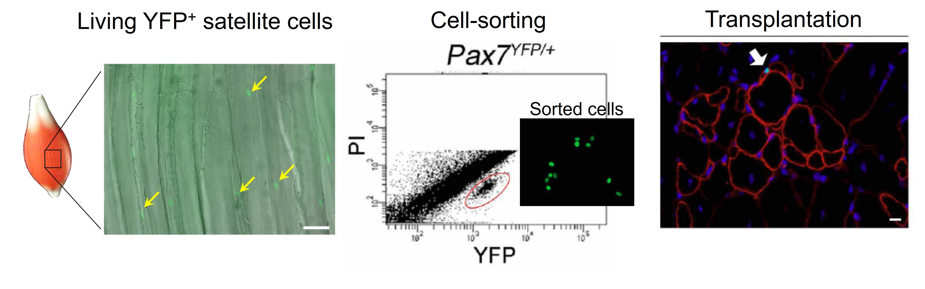
Figure 3. New tool to visualize satellite cell population
Regional specificity in skeletal muscle
Skeletal muscle is the largest tissue in the adult human body accounting for 40-50% of the body weight, composed of approximately 640 individual muscles responsible for movement and locomotion by generating contraction forces. Skeletal muscle has functionally heterogeneous properties: limb and trunk muscles function in body posture, locomotion, and respiration; while craniofacial muscles mainly control facial expression, speech, feeding activity, and eye movement.
In muscle diseases such as muscular dystrophy and sarcopenia, some muscles are particularly affected but not others, e.g. Duchenne muscular dystrophy (DMD) patients show a severe pathology in limb and trunk muscles, whereas eye muscles are totally spared. However, the underlying mechanism of the region-specific pathology is largely unknown. Several studies including ours revealed that satellite cells, between muscles and within a myofibre, are a functionally heterogeneous population in adults (Ono et al., Dev Biol 2010; Ono et al., J Cell Sci 2012; Kitajima et al., Method Mol Biol 2016). This heterogeneity may be linked to the body-region-specific pathological phenotypes in muscular diseases. Our laboratory is interested in understanding the cellular and molecular mechanisms that regulate the satellite cell heterogeneity and the relationship between the region-specific muscle pathology and functional heterogeneity of satellite cells. We are investigating 1) when the region specificity (positional memory) is established, 2) how it is maintained, and 3) what function(s) it fulfills in adult muscle.
Molecular mechanism of muscle plasticity
Skeletal muscle is a highly plastic tissue that functionally adapts its structure and metabolism in response to chemical and mechanical stimuli. Moderate endurance training mainly induces adaptation of oxidative slow-twitch fibers, while high intensity resistance training changes contractile and metabolic profiles of glycolytic fast twitch-fibers. Conversely, muscle atrophy is induced by mechanical unloading such as bed-rest, immobilization, and disuse. Our laboratory is interested in understating molecular mechanisms that regulate the muscle plasticity (Seko et al. FASEB J 2015; Kitajima and Ono, J Endocrenol 2016; Kitajima et al., Nutrients 2017). We are also pursing the impact of aging on muscle plasticity for developing strategies to treat age-related sarcopenia.
Metabolic control of skeletal muscle
Skeletal muscle is a major metabolic organ that plays a pivotal role in glucose metabolism and thus has been recognized as a therapeutic target, not only for muscle-wasting diseases, but also for patients with type 2 diabetes. We recently identified the zinc finger MYND domain-containing protein 17 (Zmynd17) as a muscle specific gene predominantly expressed in fast-glycolytic muscles. Zmynd17 is a metabolic stress-inducible factor that maintains muscle mitochondrial integrity, with its deficiency profoundly affecting whole-body glucose metabolism (Fujita et al., FASEB J 2018; Yoshioka et al., Front Cell Dev Biol 2019)(Figure 4). Our laboratory is now investigating how skeletal muscle regulates systemic energy metabolism.
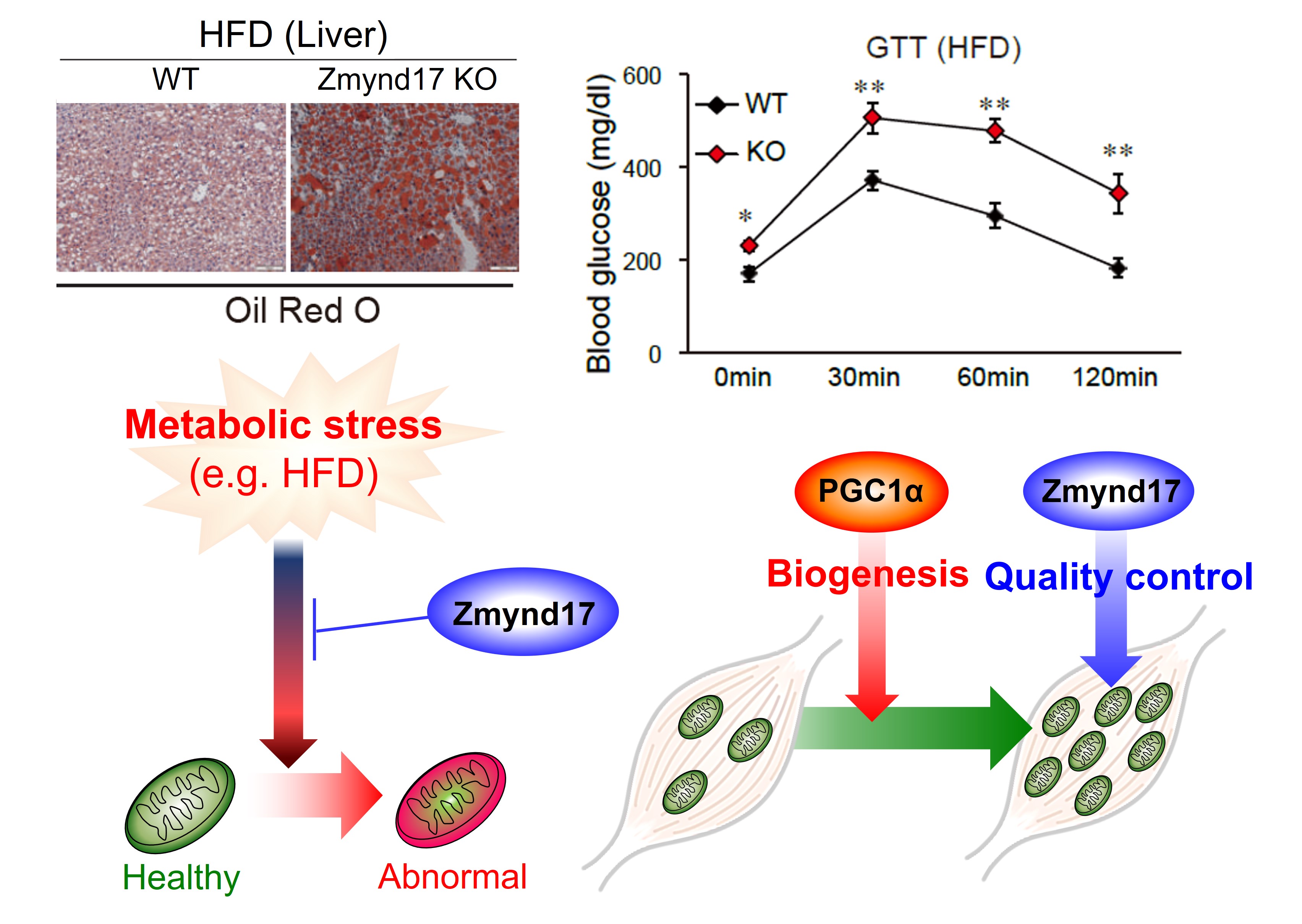
Figure 4. Zmynd17 controls mitochondrial quality